Chapter 2 · complete English translation
Keratoconus
Chapter 2: Keratoconus
- Anatomical, embryological, and physiological foundations
- The visual function of the cornea
- Biomechanics of the cornea
- Corneal ectasias
- Keratoconus
- Topography devices
- Basic indices and maps of the Sirius device
- Topographic features of keratoconus
2.1 Anatomical, Embryological, and Physiological Foundations
2.1.1 Embryological Overview
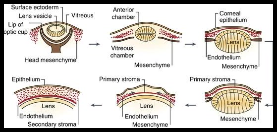
Corneal development begins embryologically with the separation of the lens vesicle from the ectoderm. At the end of the fifth week of pregnancy, the ectoderm consists of two layers of epithelial cells lying on a thin basal lamina. The separation of the lens vesicle causes the basal epithelial layer to secrete collagen fibrils and glycosaminoglycans. These fill the space between the lens and corneal epithelium and form the primitive stroma. Mesenchymal cells migrate from the margins of the optic disc along the posterior surface of the primitive stroma. The first waves of migration form the corneal endothelium, derived from neural crest cells (Figure 1; AAO, 2012; Snell & Lemp, 2013; Yanoff & Jay, 2014).
Subsequently, the primitive stroma hydrates to create space around the seventh week for the next wave of cell migration. Mesenchymal cells migrate into the stroma and differentiate into keratocytes. These secrete type I collagen fibrils and form the matrix of the mature, secondary corneal stroma. The morphological development of keratocytes in the posterior stroma progresses anteriorly. The cells produce proteoglycans and collagen fibrils arranged in successive lamellae. As the lamellae increase in length and width, the diameter and thickness of the cornea grow (AAO, 2012; Snell & Lemp, 2013; Yanoff & Jay, 2014).
In the third month of pregnancy, the endothelium in the central corneal region forms a single layer of flat cells. These lie on a discontinuous basal lamina, from which the Descemet membrane later develops. It consists of two regions: the lamina densa facing the corneal stroma and the lamina lucida adjacent to the corneal endothelium. Subsequently, the Descemet membrane organizes into a special structure, the fetal banded zone. At birth, it reaches its maximum thickness of approximately 3 µm. After birth, the posterior, non-banded portion of the Descemet membrane forms a homogeneous, fibrillar-granular substance whose thickness gradually increases with age (AAO, 2012; Snell & Lemp, 2013; Yanoff & Jay, 2014).
In the middle of the fourth month of pregnancy, the apices of adjacent corneal endothelial cells are connected by tight junctions (zonulae occludentes). This development coincides with the onset of aqueous humor production by the ciliary processes. Towards the end of the fourth month, the cell-free Bowman layer subsequently forms in the anterior stroma.
The corneal diameter is approximately 2 mm at the 12th week of pregnancy, 4.5 mm at the 17th week, and 9.3 mm at the 35th week (AAO, 2012; Snell & Lemp, 2013; Yanoff & Jay, 2014).
At birth, the horizontal corneal diameter is 8.8 mm, approximately 75–82% of the adult corneal size. The epithelium is 52 µm, the Bowman layer 12 µm, the central stroma 452 µm, the Descemet membrane 4 µm, and the endothelium 5 µm thick (AAO, 2012; Snell & Lemp, 2013; Yanoff & Jay, 2014).
The cornea continues to grow during childhood and reaches adult corneal size at approximately two years of age, with a horizontal diameter of 11.7 mm. The only structure that continues to thicken throughout life is the Descemet membrane; from birth to adulthood, its thickness increases by 6–11 µm (AAO, 2012; Snell & Lemp, 2013; Yanoff & Jay, 2014).
2.1.2 Anatomical and Physiological Overview
The cornea forms the transparent anterior sixth of the outer wall of the eyeball and acts as a non-spherical light refractor. The mean vertical corneal diameter is 10.6 mm, smaller than the horizontal diameter of 11.7 mm. The radius of curvature of the anterior surface in the central region is 7.8 mm, smaller than the radius of curvature of the sclera of 11.5 mm. Between the two lies a transition zone, the limbus, with a width of 1.5–2 mm. The radius of curvature of the posterior corneal surface is 6.5 mm. The diameter of the central optical zone is 3–4 mm.
The cornea contributes approximately 43.25 dpt (75%) to the total refractive power of the normal human eye of approximately 58.62 dpt. It is also the principal source of astigmatism in the optical system. The cornea is thinnest in the center, with a thickness of approximately 520 µm; towards the periphery, the thickness gradually increases to more than 700 µm. The corneal surface area is 1.38 cm² (Snell & Lemp, 2013).
For nutrition, the cornea depends on glucose diffusing from the aqueous humor and oxygen diffusing through the tear film. Additionally, the peripheral cornea receives oxygen from the limbal circulation (AAO, 2012).
The cornea possesses one of the highest densities of free nerve endings in human tissue. Its sensitivity is 122 times greater than that of the conjunctiva. The nerve endings originate from the long ciliary nerves and the subepithelial plexus. Neurotransmitters of the cornea include acetylcholine, catecholamines, substance P, calcitonin gene-related peptide, neuropeptide Y, vasoactive intestinal peptide, galanin, and methionine-enkephalin (AAO, 2012; Snell & Lemp, 2013).
From anterior to posterior, the cornea consists of the following layers:
1. Epithelium
The epithelium is stratified, non-keratinized, and consists of 5–7 cell layers; its thickness is 50–100 µm (AAO, 2012; Snell & Lemp, 2013; Yanoff & Jay, 2014).
- Basal cells: a layer of columnar basal cells interconnected by desmosomes and attached to the basement membrane by hemidesmosomes. The limbal basal cells produce new epithelial cells. The basement membrane is PAS-positive and firmly attached to the Bowman layer.
- Wing cells: two or three rows of wing cells connected to each other by desmosomes.
- Surface cells: two or three rows of superficial squamous epithelial cells. They contribute significantly to maintaining the smoothness of the refractive ocular surface together with the tear film anterior to the cornea. They also stabilize the precorneal tear film and form a mechanical barrier against the penetration of pathogens and tear fluid into deeper layers. This layer physiologically renews every 7–14 days. It accounts for 5–10% of the corneal thickness and is 50–70 µm thick.
2. Bowman Layer
This layer lies immediately beneath the basement membrane of the epithelium. It is 8–12 µm thick and consists of randomly arranged collagen fibers of types I and V with a diameter of approximately 30 nm. It is an acellular layer that does not regenerate after injury. It is thought to act as a barrier against growth factors secreted by epithelial cells. When these factors reach the stromal cells, they can trigger the transformation of stromal cells into fibroblasts. This explains the opacities observed after procedures that damage this layer (AAO, 2012; Snell & Lemp, 2013; Yanoff & Jay, 2014).
3. Stroma
The corneal stroma constitutes approximately 85% of the corneal thickness, similar to the dermis of the skin in this regard (AAO, 2012; Snell & Lemp, 2013; Yanoff & Jay, 2014).
- Keratocytes: 3–5% of the cells. They resemble fibroblasts, are rich in mitochondria, rough endoplasmic reticulum, and Golgi apparatus, are connected by gap junctions, and are more densely distributed in the anterior stroma. They secrete collagen fibers and proteoglycans.
- Collagen fibers: types I, III, V, and VI. They are arranged in 200–300 lamellae parallel to the corneal surface. Within each lamella, the fibers run parallel; to the fibers of the adjacent lamella, they are approximately at right angles. This organization contributes to the biomechanical properties of the cornea.
- Extracellular matrix: It consists primarily of glycosaminoglycans, predominantly keratan sulfate and chondroitin sulfate, in a ratio of 3:1. It maintains a constant distance of approximately 67 nm between the collagen fibrils.
The corneal stroma differs from other connective tissues by its transparency and its biomechanical properties. Both features are based on the precise, specialized arrangement of stromal fibers and the extracellular matrix. The network reduces light scattering and increases the tensile strength of the cornea. The peripheral stroma is thicker than the central stroma; in the periphery, the fibers can change direction and run circumferentially.
The cornea maintains its transparency through:
- the characteristic arrangement of the lamellae;
- destructive interference, since the collagen fibers are smaller than the wavelength of visible light;
- the maintenance of a limited corneal hydration of approximately 78% through the epithelial and endothelial barriers;
- the swelling pressure, generated by the repulsion of negatively charged glycosaminoglycan molecules, which counteracts the intraocular pressure.
4. Dua's Layer
A new corneal layer was discovered and named after its discoverer, the Indian scientist Harminder Dua, as Dua's layer. It lies between the corneal stroma and the Descemet membrane. Although only 15 µm thick, it possesses high tensile strength and stiffness. Its existence was confirmed by the careful injection of air bubbles into the cornea, with which the corneal layers were separated from each other and subsequently examined by electron microscopy; this enabled thousandfold magnification.
The discovery of Dua's layer may contribute to the explanation and treatment of various corneal diseases, such as the acute corneal edema that occurs in keratoconus through rupture of this layer. Furthermore, it has contributed to the advancement of lamellar keratoplasty. Its possible role in glaucoma remains a subject of research, as it transitions into the lamellae of the trabecular meshwork (Dua, Faraj, & Said, 2015).
5. Descemet Membrane
The Descemet membrane forms the basement membrane of the corneal endothelium. It consists predominantly of type IV collagen and is divided into two regions: an anterior banded and a posterior non-banded portion. Its thickness increases from 3 µm at birth to 10–12 µm in adulthood, as the endothelial cells of the amorphous, non-banded zone gradually add additional posterior layers (AAO, 2012; Snell & Lemp, 2013; Yanoff & Jay, 2014).
6. Endothelium
The endothelium consists of densely packed, interlocking cells arranged predominantly as a mosaic of hexagons. The lateral walls of the corneal endothelial cells are highly convoluted and contain folds as well as finger-like projections that interdigitate with the projections of adjacent cells. Numerous gap junctions enable cytoplasmic transport between the endothelial cells.
At the apical portions of the lateral walls facing the anterior chamber are tight junctions. They prevent permeability for substances with a molecular weight greater than 87 kDa and reduce the passive transport of fluid and electrolytes from the anterior chamber into the corneal stroma (AAO, 2012; Snell & Lemp, 2013; Yanoff & Jay, 2014). Human corneal endothelial cells do not proliferate in vivo but can divide in cell cultures. Although some evidence suggests the presence of peripheral endothelial stem cells, cell density decreases over time. Lost cells are replaced by enlargement and spreading of adjacent cells, particularly after trauma or surgery (AAO, 2012; Snell & Lemp, 2013; Yanoff & Jay, 2014).
Cell density varies across the endothelial surface and is physiologically highest in the periphery. Normal endothelial cell density ranges from 2,000 to 3,000 cells/mm². The endothelial cells maintain corneal transparency through two functions: as a barrier to the aqueous humor and as a metabolic pump. As cell density decreases, permeability increases and the number of pump sites declines. However, the cell density at which corneal edema develops is not an absolute value. Endothelial changes can be reversible (pseudoguttata) or permanent (cornea guttata) (AAO, 2012; Snell & Lemp, 2013; Yanoff & Jay, 2014).
2.2 Visual Function of the Cornea
The cornea contributes on average 42 dpt, the largest share of the refractive power of the eye. This can be calculated using the refractive power law of a spherical surface (AAO, 2009; M. Sinjab, 2009):
P = (n2 - n1) / r
where n1 denotes the refractive index of the first medium, n2 the refractive index of the second medium, and r the radius of curvature of the spherical surface. For air, the refractive index is 1; for the tear film and aqueous humor, 1.336; and for the cornea, 1.376. With a mean radius of curvature of the anterior corneal surface of 7.8 mm and the posterior surface of 6.5 mm, the following values are obtained:
- Refractive power of the air/tear film interface: 43.1 dpt;
- Refractive power of the tear film/cornea interface: 5.1 dpt;
- Refractive power of the cornea/aqueous humor interface: -6.2 dpt.
Since the total refractive power of the cornea corresponds to the sum of the refractive powers at the interfaces, the mean refractive power of the cornea is approximately 42 dpt. The interface between air and the tear film thus has the highest refractive power in the eye. Therefore, tear film disturbances lead to blurred vision and deterioration of visual quality.
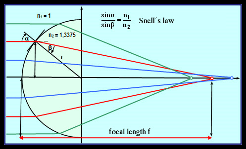
The corneal refractive power can also be calculated using ray tracing and Snell's law (Figure 2). This technique is used by topography devices to generate a refraction map:
n1/n2 = sin α2 / sin α1
n1 and n2 are the refractive indices of the first and second medium, respectively; α1 denotes the angle of incidence and α2 the angle of refraction.
The cornea normally has a prolate shape. This means that the central region is more steeply curved than the periphery, and its refractive power decreases to reduce spherical aberration. There is also the oblate form, in which the central region is less curved than the periphery. This is one of the causes of spherical aberration that some individuals experience (AAO, 2009; M. Sinjab, 2009).
2.3 Biomechanics of the Cornea
The study of corneal biomechanics is gaining increasing importance in refractive surgery. To understand these properties, elasticity, viscosity, and viscoelasticity must be distinguished (Mazen M. Sinjab, 2011b).
- Elasticity: the ability of a tissue to return to its original shape after a deformation caused by pressure or tension.
- Viscosity: the resistance of a fluid to shear forces.
- Viscoelasticity: the deformation of a tissue under sustained pressure; after removal of the force, the tissue slowly returns to its original shape.
The cornea is a viscoelastic tissue, as it essentially consists of collagen fibers responsible for elasticity and a matrix responsible for the viscous properties (Mazen M. Sinjab, 2011b).
S. 32–43 - Biomechanics, Ectasias, and Keratoconus
The Bowman layer consists of randomly arranged type I collagen fibers and is harder and stiffer than the underlying stroma. The anterior, interwoven stroma is harder and stiffer than the posterior stroma; clinically, this is reflected in the fact that blunt dissection through the anterior stroma is more difficult than through the posterior stroma (AAO, 2012).
The viscoelasticity of corneal tissue explains some previously poorly understood phenomena, such as the ectasia triggered after refractive surgery (LASIK) and the undercorrection after myopia correction (Mazen M. Sinjab, 2011b). Tissue weakness of the cornea also influences its biomechanics and can — together with a triggering factor such as chronic eye rubbing or refractive surgery — lead to the development of corneal ectasia (Mazen M. Sinjab, 2011b).
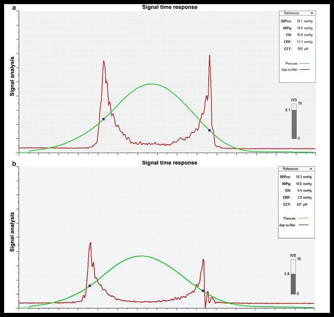
Corneal biomechanics can be assessed, for example, by measuring corneal hysteresis using an Ocular Response Analyzer (ORA; Reichert). It was observed that normal corneas have higher hysteresis than keratoconic corneas (Figure 3; Mazen M. Sinjab, 2011b). Among the newer devices for measuring hysteresis is the Corvis-ST (Oculus Optikgeräte, Germany), which will be discussed in detail later.
2.4 Ectatic Corneal Diseases and Keratoconus
Ectatic corneal defects comprise a group of conditions that alter the shape of the cornea. The cornea becomes thinner and gradually loses its tensile strength; this leads to warpage and irregular astigmatism. Maintaining an appropriate corneal shape is essential for good vision. Ectatic diseases can therefore cause significant visual impairment (Copeland & Afshari, 2013; Coster, 2002a).
These conditions are typically bilateral, confined to the cornea, and non-inflammatory. They include keratoconus, pellucid marginal degeneration, and keratoglobus. These conditions may represent a spectrum of the same disease. Also included are Terrien's marginal degeneration and post-refractive surgery ectasia (Copeland & Afshari, 2013; Coster, 2002a).
2.4.1 Keratoconus
Keratoconus is the most common ectatic corneal disease and will be discussed in detail later.
2.4.2 Pellucid Marginal Degeneration
Pellucid marginal degeneration is a relatively rare, non-hereditary, and bilateral condition that affects men and women equally. It presents as a clear, peripheral, inferior thinning of the cornea without inflammation. The diagnosis is made in most patients between the 20th and 40th year of life. Visual impairment is associated with high and irregular astigmatism. Many studies consider this condition a form of keratoconus (Sahu & Raizada, 2020).
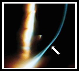
The cornea bulges anteriorly above the band of thinning (Figure 4), whereas in keratoconus the ectasia is located at the site of maximal thinning. There is no neovascularization or lipid deposition; however, scarring within the posterior stroma in the area of thinning has been observed. Rarely, acute hydrops and spontaneous corneal perforation have been reported (Sahu & Raizada, 2020).
In early cases, treatment involves contact lenses, whose fitting is more difficult in pellucid marginal degeneration than in keratoconus. In moderate cases, intrastromal ring implantation may be used. Corneal cross-linking is fundamental due to the continuous progression of the disease. In severe cases, penetrating keratoplasty (PK) remains the treatment; it is technically challenging. Wedge resection and crescentic lamellar keratoplasty are alternative or supplementary options (KUMAR, 2020).
2.4.3 Keratoglobus
Keratoglobus is a very rare, non-inflammatory, and non-hereditary condition. Unlike keratoconus and pellucid marginal degeneration, it manifests at birth, but has also been described as an acquired condition in patients with Graves' disease. It is strongly associated with conditions such as blue sclera and Ehlers-Danlos syndrome type VI (Wallang & Das, 2013).
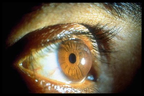
The entire cornea is markedly thinned; simultaneously, the anterior chamber depth and corneal diameters are increased (Figure 5). Keratoglobus resembles keratoconus but tends to show a spherical rather than a conical ectasia. The maximal thinning is peripheral, whereas in keratoconus it is at or near the apex of the ectasia. Vogt striae and a Fleischer ring, as seen in keratoconus, are absent in keratoglobus. Furthermore, corneal edema occurs frequently, and spontaneous perforations are seen in a high proportion of patients already at a young age (Wallang & Das, 2013).
Treatment includes spectacles for correction of high myopia and prevention of amblyopia, as well as eye protection from trauma. As a sight-preserving procedure, a lamellar tectonic graft followed by penetrating keratoplasty may be considered. The prognosis of penetrating keratoplasty in this condition is worse than in keratoconus (Wallang & Das, 2013).
2.4.4 Terrien Marginal Dystrophy
This rare condition differs from the previously described conditions but also leads to irregular corneal astigmatism. The cause is an atrophic process that weakens the support provided by the corneal periphery to the central region (Ruutila et al., 2020).
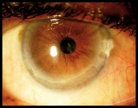
Clinical manifestations include reduced visual acuity due to unstable astigmatism. The condition typically occurs in middle age. The peripheral cornea becomes thin and atrophic; neovascularization and lipid deposits are added (Figure 6). Usually, both eyes are asymmetrically affected. Severe inflammatory exacerbations are rare; clinically, the eyes appear non-inflammatory in most cases, although signs of inflammation may be visible on slit-lamp examination. This suggests that some cases represent a mild form of Mooren's ulcer. In Mooren's ulcer, there is peripheral destruction of the cornea with ulceration and prominent inflammation. Both conditions may be endpoints of a shared disease spectrum (Chen et al., 2020).
2.4.5 Ectasia After Refractive Surgery
Corneal ectasia is a late complication of LASIK. The main causes include an undiagnosed (subclinical) keratoconic cornea with topographic signs, forme fruste keratoconus, or a suspect cornea with topographic changes — with or without definite keratoconus in the fellow eye — respectively, a structurally weak normal cornea (Santhiago, Giacomin, Smadja, & Bechara, 2016).
A careful search for risk factors is important to reduce the risk of this complication after surgery. These factors include typical topographic changes of keratoconus, which will be explained in detail later, as well as an elevated percentage of tissue ablation (PTA). The PTA is the ratio of flap thickness and laser-ablated tissue to the total corneal thickness and should be less than 40%. The residual stromal bed (RSB) should be at least 250 µm, but is less significant than the previous value. Additional factors include a central corneal thickness (CCT) below 480 µm, high myopia, young age (the latter two factors are controversial), chronic eye rubbing, a family history of keratoconus, an unstable refraction, and a corrected visual acuity of less than 1.0.
When risk factors for corneal ectasia are present, the refractive surgical method must be modified or the procedure abandoned. Additional options include flapless procedures such as photorefractive keratectomy (PRK) or the implantation of phakic intraocular lenses (Santhiago et al., 2016).
After the development of ectasia, treatment options are limited. In early stages, rigid gas-permeable contact lenses can yield good results. In moderate cases, intrastromal rings and corneal cross-linking are among the modern solutions. In advanced cases, lamellar or penetrating keratoplasty (PK) remains possible (Santhiago et al., 2016).
2.5 Keratoconus
2.5.1 Definition
Keratoconus is a bilateral, non-inflammatory, and progressive ectatic corneal disease. The ectasia describes the thinning and weakening of the cornea resulting from damage to collagen fibers in the stroma, changes in the ground substance, or reduced thickness of the normal stroma. These changes affect the central or paracentral region. The intraocular pressure thereby leads to a conical bulging of the cornea.
Usually, one eye is initially affected and later — with a variable delay — the fellow eye. In some patients, the condition remains unilateral. In Western populations, the incidence ranges from 50 to 200 cases per 100,000 persons (Kanski & Bowling, 2015; Mazen M. Sinjab, 2011b).
2.5.2 Clinical Course
Patients report reduced visual acuity due to increasing myopia and increasing astigmatism, frequent changes of glasses, or contact lens intolerance around puberty. The refraction of the two eyes is usually asymmetric.
2.5.3 Clinical Signs (Mazen M. Sinjab, 2011b)
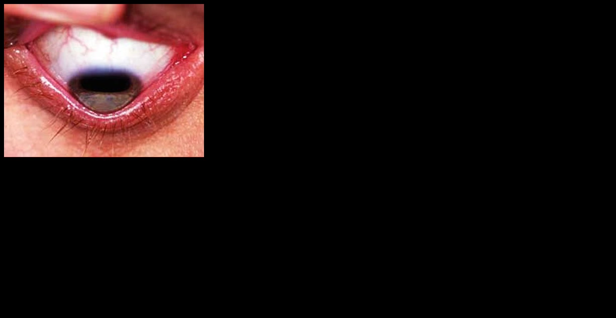
The most important clinical signs of keratoconus are summarized below (Figure 7).
Early signs:
- central or paracentral thinning of the corneal stroma; a persistent and constant sign in all cases;
- Vogt striae: fine vertical lines in the deep stroma parallel to the steepest axis; they disappear with external pressure on the cornea;
- oil drop sign on direct ophthalmoscopy;
- scissoring reflex on retinoscopy. It is a diagnostic sign of keratoconus and — in addition to other forms of irregular astigmatism — is more visible with a dilated pupil. It also has high sensitivity even for mild keratoconus forms;
- Rizzuti sign: when a narrow light beam is directed at the temporal corneal region, a reflection appears on the nasal anterior sclera. The cause is total reflection due to the optical properties of the cone;
- protrusion of corneal nerves, a less important sign.
Late signs:
- irregular myopic astigmatism. As thinning progresses, the cornea becomes thinner and visual acuity continues to decline;
- Munson sign: V-shaped bulging of the lower lid on downward gaze, because the cone pushes the lid outward;
- Fleischer ring: deposition of iron granules in the basal epithelial cells that partially or completely encircle the base of the cone.
Signs in advanced stages:
- Acute hydrops: usually a spontaneous rupture of the Descemet membrane occurs in the central region. Aqueous humor then enters the cornea and causes edema with sudden visual deterioration. The rupture heals spontaneously within 6–12 weeks and leaves a scar in the deep stroma. A peripheral rupture may have less impact on vision. Hydrops is treated conservatively with hyperosmolar agents, patching, or long-term with a contact lens; bandage contact lenses may be added for pain relief. Surgery should not be decided upon prematurely.
2.5.4 Associated Conditions in Keratoconus
Systemic associated conditions (Coster, 2002b; Kanski & Bowling, 2015): Down syndrome (trisomy 21), Turner syndrome, Ehlers-Danlos syndrome, atopy, mitral valve prolapse, and Marfan syndrome.
Ocular associated conditions: vernal keratoconjunctivitis, blue sclera, ectopia lentis, retinitis pigmentosa, ill-fitting contact lenses, chronic eye rubbing (possibly a mechanical trigger of keratoconus), and aniridia.
2.5.5 Heredity
Heredity plays a role in a subset of cases, but its pattern is not definitively known. Studies report a disease rate of 6–19% in first-degree relatives; topographic abnormalities are also more frequently found in these relatives. The disease rate in offspring does not exceed 10% (Nowak & Gajecka, 2011).
Chromosomes 16 and 20 have been proposed as possible responsible gene loci; chromosome 21 is also relevant due to its close association with Down syndrome. The reported prevalence of this association is 0.5–15% (Nowak & Gajecka, 2011). More recent studies found changes in the ZNF469 gene in 12% of individuals with keratoconus (Lechner et al., 2014).
2.5.6 Classification
Several classifications of keratoconus exist. One of the most well-known is the Amsler-Krumeich classification with four stages. The stage is determined by the presence of at least one of the features listed in Table 1.
Table 1: Krumeich Classification of Keratoconus (Naderan, Jahanrad, & Balali, 2017).
| Stage | Mean Sim-K | Thickness | SE | Scar |
|---|---|---|---|---|
| I | <48 | >500 | <-5 | - |
| II | <53 | 401–500 | -5 to -8 | - |
| III | 53–55 | 300–400 | >-8 | - |
| IV | >55 | <200 | - | + |
2.5.7 Forme Fruste, Subclinical, and Suspect Keratoconus
There are differences between studies regarding definitions and diagnostic criteria. The most widely accepted definitions are as follows (Henriquez, Hadid, & Izquierdo, 2020; Klyce, 2009):
- Forme fruste keratoconus: a completely normal cornea without clinical or topographic risk factors for the development of keratoconus; the fellow eye is keratoconic.
- Subclinical keratoconus: a non-normal cornea with a topographic defect but without clear clinical signs; the fellow eye is keratoconic.
- Suspect keratoconus: a cornea with topographic defects without clear clinical signs of keratoconus; the fellow eye is either healthy or shows similar topographic changes.
2.5.8 Pellucid-like Keratoconus
This refers to a keratoconus whose topography resembles pellucid marginal degeneration. However, the thickness map presents an exception: the thinning is not peripheral and does not show the typical bell-shaped configuration (Mazen M. Sinjab & Youssef, 2012).
S. 44–75 - Topography Devices, Sirius Maps, and Artificial Intelligence
2.6 Topographers and Tomographers
Corneal topography is fundamental for obtaining information about the corneal surface. It is of great importance for the diagnosis and treatment of ectatic corneal diseases, particularly in early, subclinical, and suspect stages. It is also important for screening individuals before refractive surgery and for selecting appropriate contact lenses.
Topographic imaging is based on several techniques that have evolved over time. Initially, the Placido disc and computerized videokeratoscopy were used, providing information about the anterior corneal surface. The subsequent Scheimpflug technique overcame a weakness of the earlier method by capturing the posterior surface, providing additional information with less influence from eye movements. Finally, OCT-based imaging was introduced; it overcomes further limitations and enables more precise depiction of scars.
2.6.1 Keratometer and Photokeratoscope
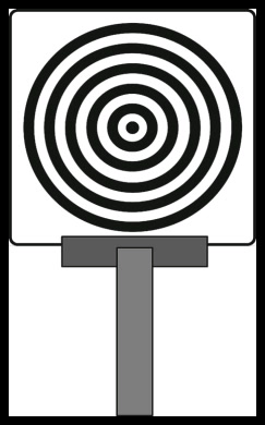
The Placido disc is an inexpensive, handheld instrument with a central aperture and nine concentric rings that alternate between light and dark (Figure 8). The examiner observes through the aperture the mirror image of the rings on the anterior corneal surface corresponding to the first Purkinje image. The pattern of ring reflections is then examined for irregular astigmatism or keratoconus.
The photokeratoscope operates on the same principle but has a rear illumination and a camera that captures an image of the ring reflection pattern (Figure 9). The camera is positioned where the examiner's eye was previously. The technique provides a topographic record of 55–80% of the total corneal circumference but inadequately or not at all captures the central region (the central 3-mm zone).
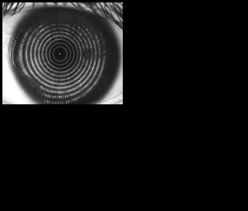
2.6.2 Computerized Videokeratoscopy
Moderate and severe keratoconus forms are easily diagnosed with the aforementioned methods. Mild and central forms, where ring crowding is minimal, are more difficult to detect. Therefore, modern imaging techniques were developed that process images with a computer and produce precise color-coded digital maps. One such light-projection-based device consists of a Placido disc and a central camera and can assess the central and paracentral corneal region.
2.6.3 Orbscan Topographer
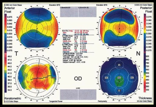
The Orbscan combines the Placido disc with an optical scanning slit to examine the anterior and posterior corneal surfaces. In less than two seconds, the device captures 7,000–9,000 points. From these, several maps are generated (Figure 10), including curvature, thickness, and anterior and posterior elevation maps. A weakness is the influence of corneal scars: the measured thickness appears lower than actual and the elevation map may mimic the pattern of an ectatic disease (Oliveira, Ribeiro, Franco, & Optometry, 2011).
2.6.4 Pentacam
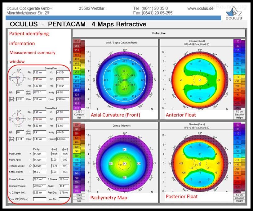
The Pentacam uses a rotating Scheimpflug camera with a 475 nm LED light source. The technique can image the anterior segment from the anterior surface of the cornea to the posterior surface of the lens, provided the pupil is sufficiently dilated. Within two seconds, 50 cross-sectional images of a complete corneal cross-section from limbus to limbus at angles from 0 to 180° are captured. The images overlap in the central region; their combination produces a complete image with 25,000 real points (138,000 in the HR version) (OCULUS).
Unlike the Orbscan, the Pentacam images contain common points (Figure 11); the Orbscan captures separate vertical cross-sections. The Pentacam can therefore compensate for eye movements through reconstruction of the central points. Moreover, it provides more information about the cornea and anterior segment, including the anterior chamber angle and the iris. This is particularly important, for example, in planning refractive procedures such as the implantation of phakic intraocular lenses (Mazen M. Sinjab, 2018b).
2.6.5 Sirius
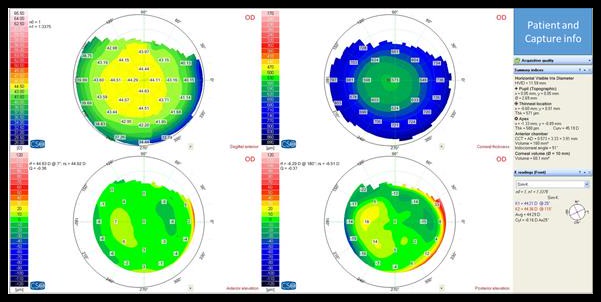
The Sirius device combines the Placido disc with a rotating camera. Twenty-five images are captured and analyzed per second. More than 21,000 points of the anterior surface and 16,000 points of the posterior surface are obtained (CSO, 2018).
The device provides comprehensive information about the anterior and posterior corneal surfaces (curvature and elevation) as well as corneal thickness (Figure 12). Additionally, it offers aberration analysis, glaucoma analysis (thickness-adjusted pressure and anterior chamber angle), and a specialized keratoconus analysis based on a support vector machine algorithm, a method of artificial intelligence (Arbelaez et al., 2012; CSO, 2018).
The device has been in the Department of Ophthalmology and Ophthalmic Surgery at Al-Mouassat University Hospital since 2010. It has a large image archive; data obtained from this archive are used in the present study.
2.6.6 MS 39 as an Example of AS-OCT
The MS 39 combines the Placido disc with anterior segment optical coherence tomography (AS-OCT). Twenty-five images are captured and analyzed; more than 31,000 points of the anterior surface and 25,000 points of the posterior surface are obtained (CSO).
In addition to the information provided by the Sirius, the device offers epithelial thickness data, which helps in detecting early keratoconus forms. It can also image the cornea in the presence of scars and provide accurate quantitative measurements of the anterior chamber angle and the corneoscleral junction (CSO). This technique is currently considered the most advanced in topographic imaging, as it overcomes the limitations of earlier methods (Figure 13).
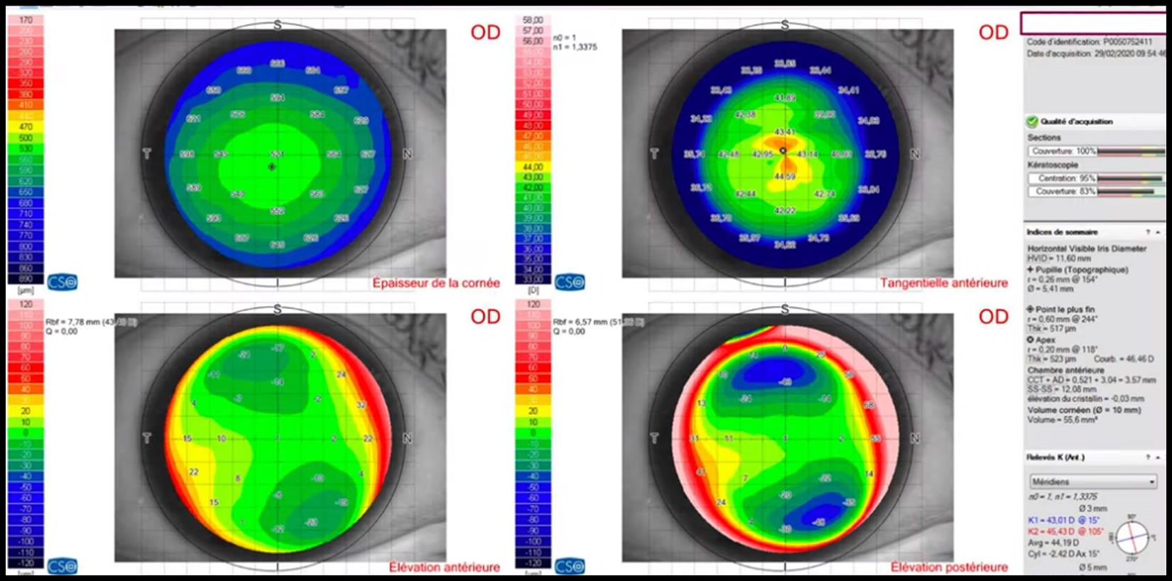
2.7 Basic Maps and Indices of the Sirius Device (CSO, 2018)
2.7.1 Basic Maps
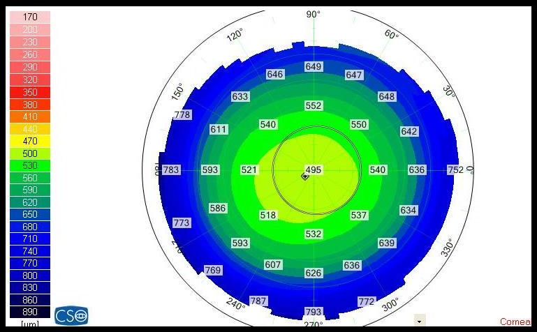
1. Corneal Thickness Map: The thickness is displayed in micrometers. Areas with greater thickness appear in cool colors (blue-green), thin areas in yellow-red (Figure 14). The computer measures corneal thickness at each point based on the elevation maps.
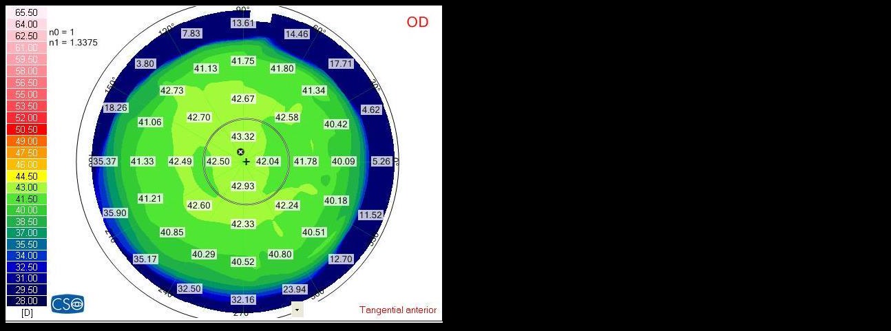
2. Anterior Tangential Map: The curvature power is given in millimeters or diopters (Figure 15). This map is the most sensitive to local curvature changes, is relatively resistant to poor fixation during image acquisition, and is better suited for assessing the corneal periphery and its refractive power. It is based on the principle of circles tangent to the corneal surface at the measurement point; the radius of curvature at this point corresponds to the radius of the tangential circle (Figure 16):
P = (n2 - n1) / r
where n1 is the refractive index of the first medium, n2 is the refractive index of the second medium, and r is the radius of curvature of the spherical surface.
3. Posterior Tangential Map: The curvature power is displayed in millimeters or diopters and calculated by the same method as for the anterior surface.
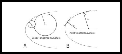
4. Anterior Sagittal Map: The curvature power is displayed in millimeters or diopters (Figure 17). For each point, a tangent to the corneal surface is drawn at measurement point A. The normal to this tangent intersects the reference axis — the normal axis to the projection of the Placido disc — at point B. The distance AB is therefore the radius of curvature at point A. Using the equation above, the refractive power is calculated; the values are positive (Figure 16).
5. Posterior Sagittal Map: Here too, the curvature power is displayed in millimeters or diopters (Figure 17) and calculated as for the anterior surface. The values are negative because the posterior corneal surface acts as a concave lens.
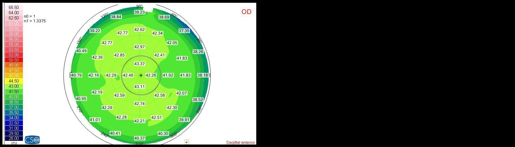
6. Anterior Elevation Map: Elevation maps are more accurate than curvature maps for assessing both corneal surfaces because they are less influenced by tear film disturbances and contact lenses. The maps are generated based on a reference surface. Points above the reference surface are considered elevations and are given in positive micrometer values; points below are depressions with negative values. Points of contact between the reference and the examined corneal surface have a value of zero.
The computer proposes an individual reference surface for each captured cornea:
- Spherical: best suited for showing details of an irregular cornea and the location of a cone;
- Aspherical: serves to display the actual corneal shape;
- Aspheriotoric (Toric Ellipsoid): an ellipsoid whose cross-section is elliptical rather than circular. This shape is particularly well suited for approximating an uneven corneal surface, as in astigmatism;
- Float: the sum of elevations and depressions around the reference surface is equal; this method can be combined with the previously mentioned reference surfaces.
7. Posterior Elevation Map: It is also based on a reference surface; the elevation values are given in micrometers (Figure 18).
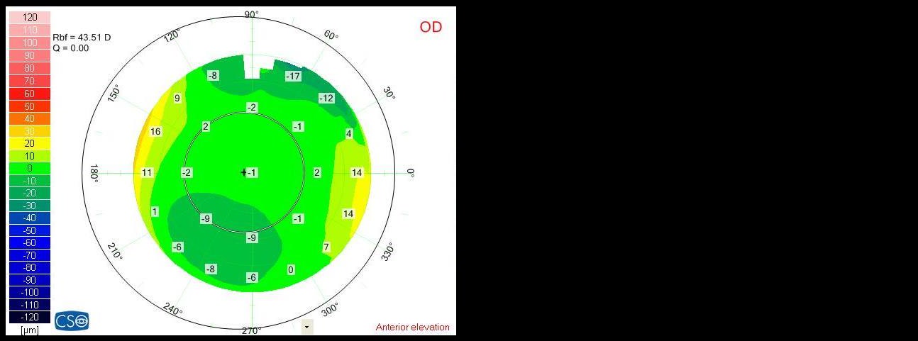
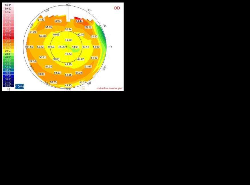
8. Anterior Refraction Map: The refractive power is given in diopters and calculated with the stromal refractive index (1.376). The map assesses the optical power of the examined surface. It uses focal lengths rather than pure curvature values and accounts for spherical aberrations. The corneal refractive power is greater in the periphery than in the center because the peripheral and central focal lengths differ (Figure 19).
9. Posterior Refraction Map: The refractive power is given in diopters and calculated using the stromal refractive index (1.1376) and the aqueous humor (1.1336); at each point, Snell's law is applied (Figure 19).
10. Equivalent Refraction Map: The refractive power is given in diopters, calculated with the stromal refractive index (1.1376), and determined for each point according to Snell's law.
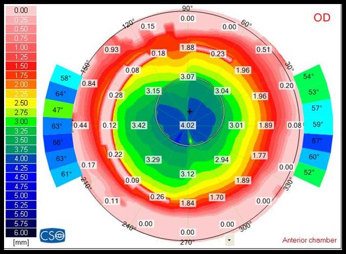
11. Anterior Chamber Map: It shows the depth of the anterior chamber and the angle between the cornea and iris (Figure 20).
12. Background Map: It shows the position of the patient's eye during image acquisition. Note: The previously mentioned maps can be displayed in three dimensions.
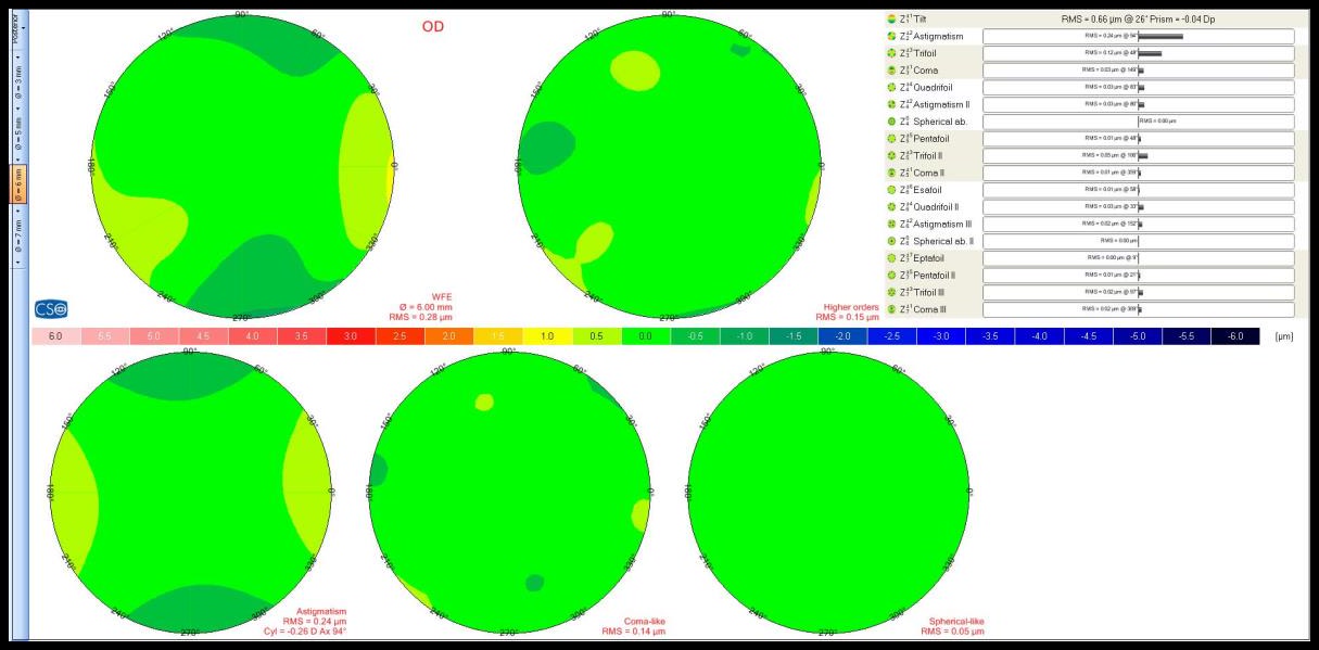
13. Zernike Analysis: The Zernike analysis describes the wavefront and its deformations as light passes through refractive media, i.e., the aberrations. The lower the aberrations, the better the optical quality of the system. The analysis shows both the total amount of aberrations and the individual aberration types. The Zernike pyramid includes 36 orders; the aberrations of the examined cornea are displayed as magnitude and form (Figure 21).
2.7.2 Basic Indices (Arbelaez et al., 2012)
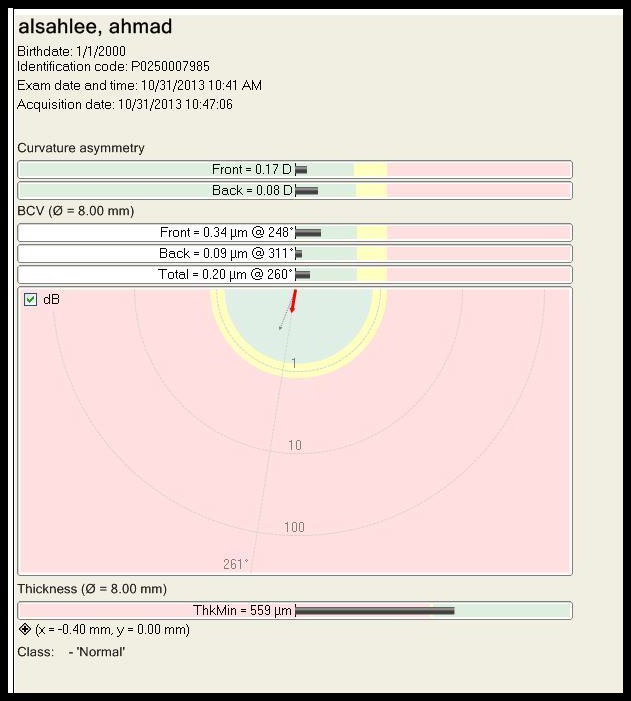
Advanced keratoconus stages are easily diagnosed from topographic maps. Early stages and suspect cases are more difficult. Many studies therefore attempted to develop different indices for objective diagnosis. The CSO device uses the following indices for classifying various keratoconus stages and suspect cases (Figure 22): abnormal cornea (Abnormal), keratoconus-compatible cornea (Keratoconus Compatible), subclinical keratoconus (Subclinical Keratoconus), and normal cornea (Normal).
Curvature-based indices:
- Anterior corneal curvature symmetry index (SIf): difference of the mean anterior tangential curvature of two upper and lower circular areas with a radius of 1.5 mm. Positive values mean that the lower half is more steeply curved; negative values mean that the upper half is more steeply curved.
- Posterior corneal curvature symmetry index (SIb): difference of the mean tangential curvature of two upper and lower circular areas with a radius of 1.5 mm. SIb is given in diopters. Because of the difference between the refractive index of the cornea and that of the aqueous humor, the values are negative compared to SIf.
Elevation-based indices: Within a diameter of 8 mm, an aspheriotoric best-fit reference surface is used. The data are decomposed into seven orders of Zernike polynomials centered on the corneal apex.
- Anterior corneal surface best-fit radius (Rbf f): the apical radius in millimeters of a best-fit ellipsoid adjusted to the eccentricity of the normal eye within the 8-mm circle.
- Baiocchi-Calossi-Versaci index of the anterior and posterior surfaces (BCVf and BCVb): The BCV is calculated in micrometers from the aberrations of the anterior and posterior corneal surfaces most strongly associated with the diagnosis of keratoconus: vertical trefoil (C3,-3), vertical coma (C3,-1), horizontal coma (C3,+1), primary spherical aberration (C4,0), second-order vertical coma (C5,-1).
- Root mean square of higher-order aberrations (RMS HOA): They are examined on the anterior and posterior corneal surfaces. The value is considered normal below 0.25 dpt, suspect between 0.25 and 0.50 dpt, and abnormal above 0.50 dpt.
Thickness-based indices: Since keratoconus is by definition a thinning of the stroma at the location of the cone, the thinnest corneal value within an 8-mm circle is used as an index to distinguish keratoconic from normal corneas.
2.8 Topographic Features of Keratoconus
Keratoconus shows topographically an abnormal anterior curvature map together with an abnormal posterior elevation map. The presence of only one of these two criteria is insufficient for the diagnosis of keratoconus; the cornea may then be classified as suspect for keratoconus (Mazen M. Sinjab, 2018d).
Corneal thickness is not an independent diagnostic criterion. A keratoconic cornea may be relatively thick and a normal cornea relatively thin. However, thin corneas (thinnest point <470 µm) are considered a risk factor in refractive surgery and are classified as suspect (Anayol et al., 2014).
2.8.1 Curvature Maps
Four patterns can be distinguished (Mazen M. Sinjab, 2018a; Figure 23):
- Symmetric: includes spherical, elliptical, and symmetric bowtie forms. They are considered abnormal when the mean curvature exceeds 47.2 dpt in Orbscan devices and 48 dpt in Pentacam devices.
- Asymmetric: includes an asymmetric bowtie with superior or inferior curvature and an isolated superior or inferior curvature. It is considered abnormal when the mean curvature value is abnormal or when there is a relevant difference between the superior (S) and inferior (I) mean within a 3-mm circle and a 30° sector (I-S >1.4 or S-I >2.5).
- Skewed: includes symmetric and asymmetric bowtie forms with skewing. It is considered abnormal when the angle between the axes of the two segments within a 3-mm circle exceeds 21° and at least 1 dpt of astigmatism is present.
- Other patterns: including crab claw, butterfly, vertical D, clown face, and vortex patterns as well as unspecific irregularity. They are always considered abnormal, except for unspecific irregularity, which occurs in 7% of the normal population.
2.8.2 Elevation Maps
The shape is assessed based on a spherical reference body. There are two main patterns (Figure 24):
- Regular pattern: isolated central island and hourglass.
- Irregular pattern: tongue-shaped form and twisted hourglass. Both patterns can occur with a large kappa angle or poor fixation.
Neither pattern alone is considered abnormal; what matters is abnormal numerical values. The values are read depending on the reference body used (8-mm diameter, float mode; S. Khachikian & Belin, 2008; S. S. Khachikian, 2012; Mazen M. Sinjab, 2018c).
For the spherical reference body, Table 2 shows the maximum threshold values of the highest point (3 standard deviations) for the anterior and posterior elevation maps:
| Elevation Map | Myopia | Hyperopia |
|---|---|---|
| Anterior surface | 7.7 | 6.5 |
| Posterior surface | 17.7 | 27.8 |
Table 2: Maximum threshold values in the elevation map with spherical reference body.
For the toric ellipsoid reference body, the maximum threshold values are given within a 5-mm circle:
| Elevation Map | Threshold within 5 mm |
|---|---|
| Anterior surface | 7.7 |
| Posterior surface | 17.7 |
Table 3: Maximum threshold values in the elevation map with toric ellipsoid reference body.